
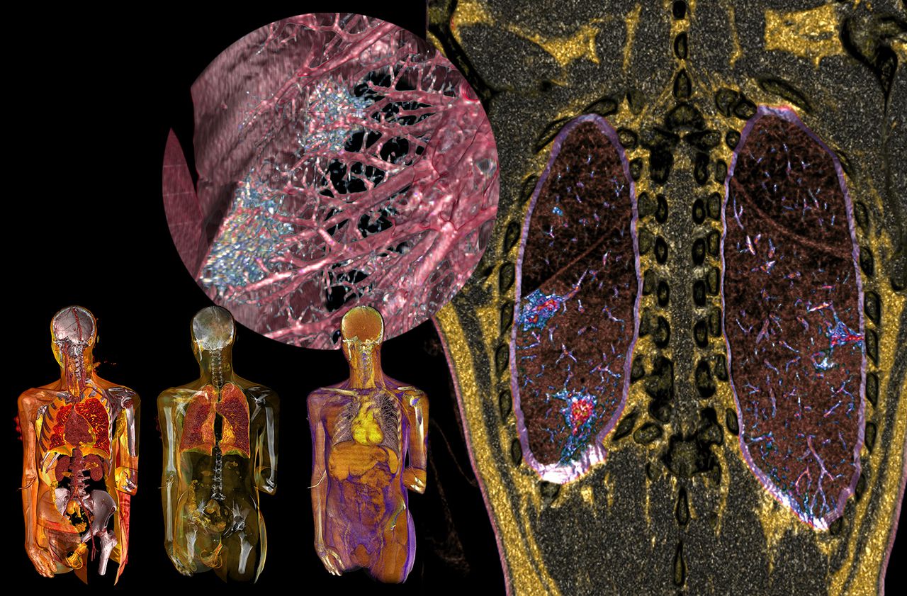
You have to go along the trachea, branch off into the lungs, cross the bronchi and bronchioles to see it. He is there, massive, motionless. Like a heap of tiny blue stones glued to each other. Under our eyes appears Sars-CoV-2 or more precisely the lesions of the disease it causes. The enemy who is said to be "invisible" therefore has a shape, a volume, a color. The pictures that scroll on the screen are the fruit of a marriage "between IT and imagery", summarizes his authorizing officer. Doctor Rodolphe Gombergh produced these chest scanners for patients infected with the virus.
Unlike conventional exams, his are coupled with high definition software (RVG-VLA), which he invented fifteen years ago to restore the volumes. They allow it to wander virtually in the body, as here in the pulmonary system damaged by the Covid-19. "It's like an interior trip in 3D, a stroll in the patient's clone - the real thing, we leave him alone," enthuses the 68-year-old radiologist.
The journey begins in its research center, boulevard Malesherbes, in Paris, where the scanner with very low radiation dose and the superpowerful computer are housed. Difficult when observing his office in a happy bazaar to imagine that he produces these images unique in the world. However, Doctor Gombergh is not a stranger in the world of medical radiology ... like art. Since the early 2000s, his work has been regularly exhibited, from the Academy of Medicine, to the Center Pompidou!
The virus is deposited on the alveoli
"The transparent man technique makes it possible to detect the smallest anomalies, that's why I wanted to apply it to the coronavirus", he says, round glasses brought back to the tip of the nose. His goal: to better understand the imprint left by this young disease which has not revealed all its secrets.
PODCAST. What are coronaviruses? Diving under the microscope in virus biology
"With this crisis, everyone is discovering how important breathing is and that the role of the lung is major", notes, for its part, Professor Chantal Raherison-Semjen, doctor at CHU Bordeaux and president of the Société de French-language pneumology (SPLF). The infected droplets will enter through the nose or mouth during one of the fifteen to twenty breaths we take per minute. They will pass the windpipe and be propelled into the bronchi.
Newsletter - The essentials of the news
Every morning, the news seen by Le ParisienI'm registering
Your email address is collected by Le Parisien to allow you to receive our news and commercial offers. Find out more
"The virus will deposit on the pulmonary alveoli, where gas exchanges and the passage of oxygen through the blood take place," says the pulmonologist. In most cases, this will cause nothing more than a small cold. But for a minority of patients, it will invade the cells and cause more or less severe inflammation. "
These so-called “frosted glass” opacities, doctors know how to recognize them on any traditional scanner. But if we observe them so distinctly on those of Rodolphe Gombergh, it is because thanks to billions of data, he was able to colorize them and give them volume. Masses of a translucent blue are as fixed in the pink vines which are the bronchioles. Some kind of virus signature.
Some pictures by a radiologist reached by the Covid
“I have seen a lot of frosted glass for many diseases, but never as much as for the coronavirus. Their extent seems linked to the seriousness of the patient's state of health, "notes the radiologist, long hair and futuristic look, who does not wear a blouse but is completely dressed in white. “The idea is to follow the behavior of these heaps of mucus, their appearance, their diffusion, their disappearance. "
The doctor scrolls through a new image. We are snorkeling in front of the blue dots that sparkle all over the lung. Impression that he is taken hostage. "On the contrary, it's not bad," he retorts. Spreading them out is a good thing, it means that the virus is starting to leave. "
He knows it all the more since this snapshot ... is that of his own infected lungs. "Yes, I'm healing," smiles the man who owes his passion for the job to his radiologist grandfather. In March, Dr. Gombergh experienced Covid symptoms. He did a first scan, the result of which was "compatible" with the disease. "For me, that doesn't mean anything: either we have something, or we don't have it. This is where I wanted to process the images with my high definition software in order to come up with something more precise. "
Imagery, a true ally
Now cured, he is convinced that the CT scan could become a tool for early detection of lesions of the disease. “With the tests, we know we have the disease. With the scan, we can see how much, ”pleads one who would like the exam to become more democratic at different stages of the infection. An opinion that is debated in the medical community.
The fact remains that imaging is a real ally for monitoring severe Covid-related damage, whether respiratory (pneumonia, respiratory distress) or vascular. Because we now know that it can cause damage to the vessels of the heart, the digestive tract, the kidneys… According to the scientific journal Nature, thromboses (clots in the vessels) occur in 20 to 30% of the most serious forms, and increase the risk of death.
"I would like us to further improve the supercomputer to reduce analysis times to a few minutes and benefit the greatest number of patients," notes Dr. Gombergh. Who knows what his next images will look like? By turning off the light, a painting shines. It was a Buddha from the Guimet museum that he was able to scan in his machines of the future! Will he expose his travels to the heart of the lungs hit by the epidemic of the century? “Maybe someday when all of this is behind us. "
VIDEO. Lungs infected with coronaviruses modeled in 3D


/cloudfront-eu-central-1.images.arcpublishing.com/prisa/S7ERVSCT4FUVX6R7TUVBDNTH5Y.jpg)


