
Prof. Sherman and other researchers were able to identify clusters of oncogenic proteins in melanoma cells • "A Step to Develop Innovative Therapies"
Another step in the fight against cancer
Photography:
Spokeswoman for the Hebrew University
Prof. Elon Sherman, a research student in his laboratory, Aran Yaakobian, and Prof. Yardena Samuels of the Weizmann Institute, were able to identify for the first time clusters of oncogenic proteins in melanoma cells, as well as observe the interaction between proteins using this method, with an accuracy of about 20 nm.
The study is another step on the way to developing innovative treatments against many types of cancer.
The healthy cells in the human body have control mechanisms (genes) that are responsible for balancing function and cellular functions.
Cancer cells are formed when there is a mutant change in the coding of certain proteins (oncogenic).
As a result, the balance given by the control mechanism is disturbed and the cancer cells perform destructive actions on the body, gaining new features such as unnecessary over-distribution.
Since cellular functions are carried out by transferring information between healthy proteins, changes in their spatial organization and interactions between them can be expected following the genetic change.
Despite this, many studies concerning the interaction between proteins in cancer cells, have found it difficult to determine how the defects in them are organized in space, and to explain the activity in the information transfer pathways during cell division.
Prof. Elon Sherman and research student Aran Yaakobian from the Institute of Physics at the Hebrew University of Israel, published a new and fascinating study regarding the spatial organization of proteins in cancer cells, in the journal "Research Cancer of the American Cancer Society AACR" - focusing on the microscopic resolution.
The method is designed to make it easier to decipher the location and organization of signaling proteins, which transmit information between cancer cells.
The method they used in their study received significant exposure in 2014, when three researchers won the Nobel Prize in Chemistry for developing the super-resolution fluorescent microscopy.
The three scientists made it possible for the first time to break the boundaries of the resolution that existed up to that time, and to look at living cell components that were only tens of nanometers in size.
Their breakthrough created a new field of optical nanoscopy (viewing extremely small sizes), which Prof. Sherman helped develop together with other researchers around the world.
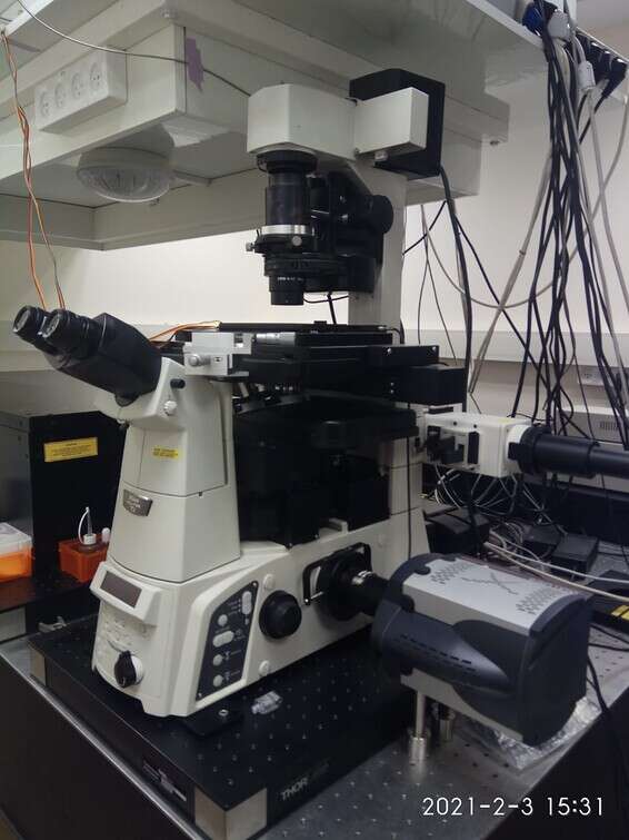
The system used by the researchers in this study is based on a Nikon microscope (eclipse Ti).
Prof. Sherman, said: "We have previously expanded the microscopic method to work in two or more colors. Why is this critical? Because only then can one look at interactions between any two proteins in a human cell and understand how signals are transmitted between cells and within cells at a single molecule resolution."
At the beginning of the present study, the researchers focused on finding the location of NRAS and BRAF, two important proteins for transmitting information between melanoma cancer cells, and even other cancers.
Currently, there is no targeted drug against mutations in NRAS, with neutralizing the action of the protein mutation being considered a particularly difficult target.
The drugs developed so far against the cells containing a mutation in BRAF are also considered less effective due to the development of the resistance of the cancer cell to the drug.
These facts place these proteins in research centers around the world for the development of drugs for cancer, with an emphasis on the development of drugs against melanoma.
Prof. Sherman, said: "We have previously expanded the microscopic method to work in two or more colors. Why is this critical? Because only then can one look at interactions between any two proteins in a human cell and understand how signals are transmitted between cells and within cells at a single molecule resolution."
He added: "The high motivation behind our study is mainly related to RAS protein, against which it is probably impossible to develop a drug. RAS is a very common oncogen in which mutations appear in up to half of certain cancers. The problem with it is that it binds the molecule with which it works so strongly. "There is no drug that can compete with it. Therefore, there is a great effort to find a solution that can work around this connection, to lead to the cure of many types of cancer."
The researchers reached a level of maximum resolution in the study, which allows targeted identification of the location of individual proteins across the mantle membrane of cancer cells, and with their help created a "statistical map" - through which can understand the cancerous spatial activity and especially how cancer cells overact and develop melanoma.
The cells formed spatial structures and fixed patterns, with the researchers being able to count the amount of proteins in each structure, characterize their motion, and identify the spatial relationships between the various proteins.
"Cancer proteins 'talk' to each other and aim to transmit a signal to cause the cell to divide rapidly and uncontrollably. They mutate, and thus are able to bypass an external signal to the cell or produce an independent signal. We think the structures we discover through the new macroscopic method control In the process of connecting the proteins and transmitting the signals, "Prof. Sherman clarified.
He added: "The clusters we uncovered were examined in previous works, but the researchers looked at them for only one protein, and could not describe the communication between the proteins. Previous methods performed were also ineffective because they involved an aggressive process of cells - torn from surfaces and looking at their remains. "The new method we used is the cleanest and most accurate that exists in the context of characterizing the files using the same innovative high-resolution microscope. You can actually count molecules in complex structures in whole cancer cells, and place them precisely high in space."
The researchers believe that information about the location and organization of key proteins in transmitting a cancer signal inside and outside the cell may in the future lead to deciphering the action of mutant cancer proteins that have undergone mutant change and science has had great difficulty understanding their function.
They are also confident that the method may pave the way for innovative and creative treatment of cancer cells.
In a future study we will be interested in examining the dynamics and mechanisms of cluster formation of proteins in cells, following the interactions of additional proteins in the signaling pathway, and investigating in depth the effect of cell resistance formation on a variety of drugs and the spatial organization of proteins.
"It is worth noting that we continue to similarly investigate signaling mechanisms in other important biological systems, such as cell activity in the immune system," the researchers conclude.