First photo in the world of the Omicron variant, created in the research area of Multimodal Medicine of the Child Jesus (coordinated by Prof. Carlo Federico Perno), under the direct supervision of Prof. Claudia Alteri (in collaboration with the State University of Milan), by Valentino Costabile, Rossana Scutari, and Luna Colagrossi. The photo shows the structure of the spike protein of the Omicron variant, on the right, and of the Delta variant, on the left, compared to the original SARS CoV-2 spike. Omicron has many more Delta mutations (already very varied), concentrated in an area that interacts with human cells. The red dots indicate the areas with very high variability, the orange ones with high variability, the yellow ones with medium variability, the green ones with low,and the celestial ones with little variability. The gray area is the one that does not vary. This does not automatically mean that these variations are more dangerous, simply that the virus has further adapted to the human species by generating another variant. "Further studies will tell us if this adaptation is neutral, less dangerous, or more dangerous", comment the researchers.
First photo of Omicron from the Bambino Gesu research group
2021-11-27T17:41:33.657Z
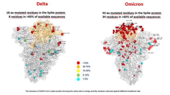
Majority mutations in area interacting with human cells (ANSA) First photo in the world of the Omicron variant, created in the research area of Multimodal Medicine of the Child Jesus (coordinated by Prof. Carlo Federico Perno), under the direct supervision of Prof. Claudia Alteri (in collaboration with the State University of Milan), by Valentino Costabile, Rossana Scutari, and Luna Colagrossi. The photo shows the structure of the spike protein of the Omicr