
Social News
Written by: Ouyang Dehao
2020-04-09 17:53
Date of last update: 2020-04-09 17:53The brain has more than 10 billion nerve cells. They are small in size and fast in activity. In the past, they lacked in-depth research on microscopy. Today, Dr. Xie Jianwen, Associate Professor of the Department of Electrical and Electronic Engineering and Director of Biomedical Engineering at the University of Hong Kong, led the team to develop two microscopic techniques that are believed to provide clues for the treatment of neurological diseases of the brain and find common diseases of the elderly, such as Alzheimer ’s The causes of Parkinson's disease.
Xie Jianwen pointed out that the volume of brain nerve cells is only 0.01 to 0.1 mm, and the activity speed is 0.001 seconds. If you can capture the activity of the cells, you can understand how the brain sends out signals to control various parts of the body and the causes of brain diseases. However, there is currently no technology that can perfectly capture brain cell activity. He continued to point out that the current common technologies, including "implanted electrode needles", "functional magnetic resonance", and "traditional fluorescent microscopes" all have different shortcomings.
For example, "implanted electrode needle" will cause damage to the brain, "functional magnetic resonance" has a low resolution, and "traditional fluorescent microscope" has a slow capture speed. Xie Jianwen led the team to develop two technologies, which have the characteristics of low trauma, high resolution and fast speed to capture the signals of brain cells.
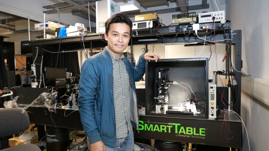
Dr. Xie Jianwen, Associate Professor of the Department of Electrical and Electronic Engineering and Director of the Biomedical Engineering Course at the University of Hong Kong, today (9th) published the research results, introducing the two newly developed micro-technology of biomedical engineering. (Provided by the University of Hong Kong)
Scanning speed is 1,000 times faster than existing laser technology
The scanning speed of the "two-photon fluorescent microscope" is 1,000 times faster than the existing laser technology. Xie Jianwen developed the "ultra-high-speed laser scanning technology", which uses parallel mirrors to allow high-speed lasers to capture the brain signals of experimental subjects, and can scan the brain 1,000 to 3,000 times per second. In terms of depth, it can also detect one-third of a millimeter below the surface of the brain, and has the advantage of not damaging the brain nerves (low trauma).
Damage to cells is reduced by at least 1,000 times
Xie Jianwen said that although the scanning speed is accelerated, the cells will be damaged after being exposed to strong light for a period of time. The "Coded Light Sheet Array Microscope" combines high efficiency and low trauma. It uses communication and broadcast technology to reorganize 3D images and capture more than 10 3D images per second, which is at least 1,000 times less harmful to cells than current technologies. , Can study cells for a long time without worrying about damage by laser.
Find out the causes of elderly diseases such as Parkinson's disease
Xie Jianwen pointed out that the two microscopic techniques can be applied in neurology and biology, for example, it can provide clues for the treatment of cerebral neurological diseases, and find out the causes of common brain diseases in the elderly, such as Alzheimer's disease and Parkinson's disease; Or observing the process of infection of cells and organs by viruses or bacteria is also helpful for studying viruses.
Xie Xu pointed out that the current team, together with nine PhD students, had received government-funded research at an early stage, and received funding from the US Institute of Health about 20 million two years ago, with a time limit of three years. He revealed that 4 million of them were invested in researching and developing microscopes, and that chemicals, wages, etc. were also expensive. He even laughed and said, "In fact, raising a white mouse is not flat."
The new 3D carbon nerve scaffold developed by the Chinese Academy of Sciences is expected to be implanted in the brain to treat brain degeneration
University of China researches the new microscope to see the application of stereoscopic cell diagnosis in the fastest 5 years
The cost of the new holographic microscope produced by the team of Nanjing University of Science and Technology is only 1% of similar products
Brain Disease, The University of Hong Kong Medical School