
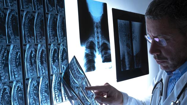
Entering the body without entering it: this is how the history of medical imaging with x-rays begins. For the first time, doctors can look inside the human body without making an incision.
Le Figaro
takes stock of the different methods.
X-rays
To carry out an x-ray, x-rays are produced by changing the path of electrons directed towards a metal plate, which then emits photons capable of passing more easily through the soft parts of the body than the hard parts, such as bone.
An image is obtained by collecting, at the exit of the organism, all the photons which have succeeded in crossing it, initially on a photographic plate but, more and more, on reception systems which make it possible to directly obtain a digital image.
The improvement of these receiving systems makes it possible to visualize very fine differences in radiation absorption and therefore very small anomalies.
It is also possible to inject
This article is for subscribers only.
You have 79% left to discover.
Subscribe: 1 € the first month
Can be canceled at any time
I ENJOY IT
Already subscribed?
Log in