
Sci-Space is the name of the new technique that will allow the creation of 4D atlases of the embryo to visualize the genes turned on in individual cells during the different stages of development: experimented on mouse embryos, it could be applied to all mammals, to study problems such as congenital malformations, developmental delays and hereditary diseases. The possibility of following the evolution of the various organs over time will also help regenerative medicine, as evidenced by the study published in Science by researchers at the University of Washington in collaboration with the Howard Hughes Medical Institute and the Brotman Baty Institute for Precision. Seattle Medicine.
Up to now, the study of the development of organs and tissues has mainly been based on the sequencing of the RNAs produced in individual cells starting from 'switched on' genes: however, most of the available techniques do not allow us to grasp the spatial context in which the cells and how it affects their development; only some methods of analysis allow to grasp this aspect, but for a limited number of genes or for small areas of tissue. Thanks to sci-Space, researchers led by Sanjay Srivatsan managed to overcome this problem by combining single-cell resolution with the spatial context of large-scale cells.
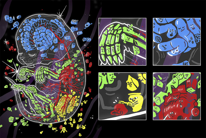
The technique, tested on mouse embryo sections on the 14th day of development, allowed us to study the activity of genes in 120,000 cell nuclei, observing how it was conditioned by the positioning of the cell in the tissue. This influence was very evident, for example, in some cartilage cells, destined to become part of the scaffolding for the bones of the skull and muzzle. The researchers were also able to study how gene expression changes during the differentiation and migration of brain cells, even discovering new 'routes' of their movements.